Sialolitíase da glândula submandibular associada a defeito ósseo de stafne: relato de caso
Clarina Louis Silva Meira2; Breno Gonçalves Daroz1; Thiago Brito Xavier1; Josiclei de Castro Moraes1; Yago dos Santos Pereira1; Diego Pacheco
Ferreira1; Célio Armando Couto da Cunha Júnior1; Hélder Antônio Rebelo Pontes1
1Federal University of Para, João de Barros Barreto University Hospital, Belem, Para, Brazil.
2Federal University of Para, Faculty of Dentistry, Belem, Para, Brazil.
DOI: 10.1900/JBPML.2022.58.429
ABSTRACT
Sialolithiasis is the formation of calcific concretions within duct of a salivary gland that affects the submandibular gland with greater prevalence. Stafne bone cavity is a rare mandibular defect of unknown etiology, which commonly presents glandular tissue inside. The aim of this work is to report the first clinical case of sialolithiasis of the submandibular gland with associated Stafne bone defect. Although most of Stafne bone cavity are associated with the salivary gland, there are no studies that prove the association between the two injuries, and further studies are needed to elucidate the relationship between the injuries.
Key words: salivary Gland Calculi; oral pathology; oral surgery.
RESUMO
A sialolitíase é a formação de cálculos no interior do ducto de uma glândula salivar que acomete com maior prevalência a glândula submandibular. O defeito ósseo de Stafne é um defeito mandibular raro de etiologia desconhecida, que comumente apresenta tecido glandular no interior. O objetivo desse trabalho é relatar o primeiro caso clínico de sialolitíase da glândula submandibular com defeito ósseo de Stafne associado.Apesar da maioria dos desfeitos ósseos de Stafne estarem associado à glândula salivar, não há estudos que comprovem a associação entre as duas lesões, sendo necessários mais estudos para elucidar a relação entre as lesões.
Palavras-chave: cálculos das glândulas salivares; patologia bucal; cirurgia bucal
INTRODUCTION
Sialolithiasis is the formation of calculi inside the duct of a major or minor salivary gland and is one of the most common diseases that affect these glands. The submandibular gland has the highest prevalence of sialolithiasis in relation to the others(1-3). This predominance can be explained by the alkaline salivary pH, which favors the precipitation of calcium salts, the thicker viscosity of the saliva due to the predominantly mucous content and the anatomical factor of the presence of the long and tortuous duct above the gland, with salivary drainage being performed against the severity(1).
Stafne’s bone defect is a rare mandibular defect of unknown etiology that appears as a round or oval unilocular radiolucency below the mandibular canal, which can occur between the mandibular angle and mandibular third molar or between the canine and mandibular premolars(4,5). The typical content of these bone cavities is salivary gland tissue, most frequently the submandibular gland. However, other types of tissue can also be found(6,7).
There are no studies in the literature reporting the association of sialolithiasis with Stafne’s bone defect. Therefore, the aim of this paper is to report the first clinical case of sialolithiasis of the submandibular gland duct with Stafne’s bone defect associated with the same side of the lesion.
CASE REPORT
A 44-year-old female patient, with a history of breast cancer with metastasis in family members,was referred to the Maxillofacial Surgery and Traumatology service of the João Barros Barreto University Hospital after consultation with an otolaryngologist reporting pain caused in the region left mouth floor.
The extraoral clinical examination showed a slight increase in ipsilateral submandibular volume with the presence of lymphadenopathy with approximately 2.5 cm in its largest diameter. Oral mucosa, with a smooth surface, firm consistency on the left mouth floor, at the height of dental elements 32 and 33 and along the path of Wharton’s duct. On occlusal radiography imaging, the presence of radiopaque foci was noted (Figure 1). The panoramic radiograph did not show an overlapping of calcifications, but an unilocular radiolucent image was shown below the mandibular canal on the left side, suggestive of Stafne’s bone defect (Figure 2).
The patient underwent surgery under local anesthesia for surgical excision of the sialolith. After suturing to prevent posterior displacement of the stone, a small superficial intraoral incision was made to expose the sialolith (Figure 3). After dissection and sufficient mobility, the sialolith was easily removed and sent for histopathological analysis. An opening for spontaneous salivary drainage was maintained (Figure 4) and, on return after one week, the sutures were in position with good healing (Figure 5). In a 1-year follow-up, the patient had good healing and no recurrence.
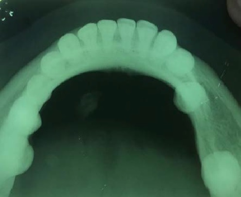
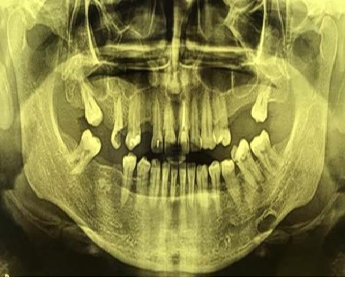
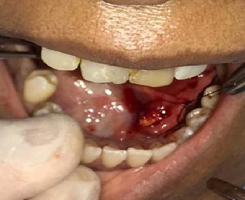
Figure 1 – Occlusal radiography showing the presence of radiopaque foci on the left side.
Figure 2 – Panoramic radiograph showing Stafne’s bone defect on the left side of the mandible.
Figure 3 – Surgical excision of the sialolith.
DISCUSSION
The submandibular gland is one of the main salivary glands, being a mixed gland and predominantly mucous, which is related to the mylohyoid muscle in the angle of the mandible(8).
Clarina Louis Silva Meira; Breno Gonçalves Daroz; Thiago Brito Xavier; Josiclei de Castro Moraes; Yago dos Santos Pereira; Diego Pacheco Ferreira; Célio Armando Couto da Cunha Júnior; Hélder Antônio Rebelo Pontes
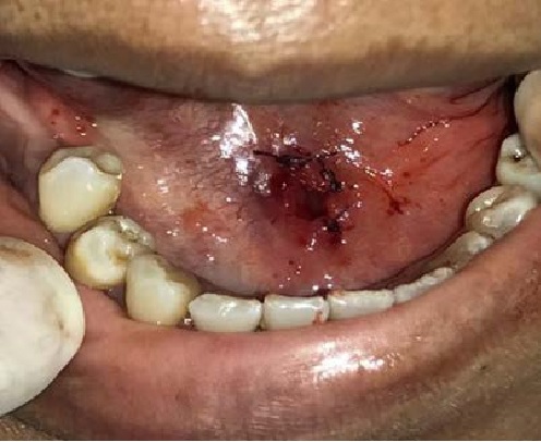
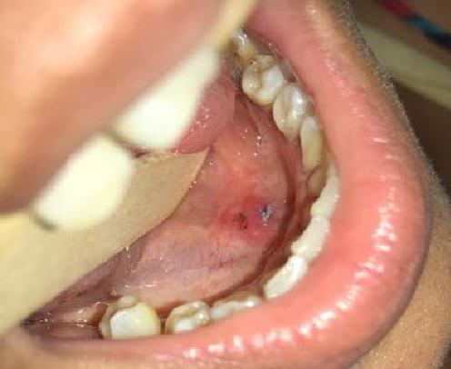
Figure 4 – Suture of the surgical incision maintaining an opening for salivary drainage.
Figure 5 – Clinical appearance of the scar at 1 week of postoperative follow-up.
In relation to the other glands among them, we can mention the more alkaline salivary pH, the higher concentration of calcium, the viscosity of the saliva produced and the characteristics of the salivary duct, which in this gland is long and tortuous, in addition to being located above it, which results in in a drainage against gravity(1).
Asialolithiasis is more prevalent in adult males and presents with painful symptoms and edema in the region of the affected gland, with reported worsening of pain during meals(1,8-10). The clinical presentation of our case corroborates these findings, despite the rarer involvement in females.
The standard radiograph for detecting submandibular and lingual salivary calculi is occlusal. However, not all calcifications can be detected by conventional radiography methods, requiring more advanced tests such as computed tomography, sialography, ultrasound, scintigraphy or magnetic resonance(11). Despite the fact that tomography has already been identified as a more sensitive and specific method for diagnosis, it was not feasible to use it in the case described, due to the patient’s socioeconomic limitations(10). However, occlusal radiography and clinical findings were sufficient to confirm the diagnosis and plan a minimally invasive surgery.
Smaller salivary calculi that do not cause obstruction in the duct has physical therapy as an alternative to non-surgical treatment, whereas larger and symptomatic calculi that obstruct salivary drainage must be surgically removed(10). The surgical approach is according to the location of the calculus in relation to the gland or duct. When the sialolith is in the anterior or middle third of the duct, removal can be done by surgically exploring the duct, while the more posterior location of the sialolith may require a sialoadenectomy(11). Recent advances propose a minimally invasive treatment, eliminating the need for sialoadenectomy, removing the sialoliths through endoscopic procedures or intraoral surgical procedures(12). Minimally invasive treatment poses less risk to the structure of the salivary gland, preserving its function(9).
Stafne’s bone defect is rare and its etiology is unknown, however, the theory that the submandibular gland exerts pressure on the lingual cortex of the mandible is widely accepted(6,7). In our clinical case, the defect was diagnosed based on characteristic radiographic findings(5,6). Biopsy becomes necessary when these defects appear in anterior regions of the mandible, where they can mimic other lesions(13). The bone defect associated with an adjacent salivary gland disorder.
Despite the ipsilateral occurrence of the lesions and the fact that most of Stafne’s bone defects are associated with the salivary gland, there are no studies in the literature that prove the association between the two lesions, and it is not possible to state that there is in fact an interaction. Therefore, we believe that this is the first report of the presence of these two concomitant pathologies in the same patient. It is noteworthy that further clinical and laboratory studies are needed to elucidate the relationship between these lesions.
Conflict of Interest: All authors disclaim any conflict of interest.
Sialolithiasis of the submandibular gland associated with stafne bone defect: case report
REFERENCES
- Gulati U, Kshirsagar R, Singh G, et al. Submandibular sialolithiasis: a brief overview and report of two cases. Modern Res Dent. 2018; 1(5): 1-7.
- Holden AM, Man CB, Samani M, et al. Audit of minimally-invasive surgery for submandibular sialolithiasis. Br J Oral Maxillofac Surg. 2019; 57(6): 582-6.
- Kolomiiets SV, Udaltsova KO, Khmil TA, et al. Difficulties in diagnosis of sialolithiasis: A case series. Bull Tokyo Dent Coll. 2018; 59(1): 53-8.
- Stafne EC. Bone cavities situated near the angle of the mandible. J Am Dent Assoc. 1942; 29: 1969-72.
- He J, Wang J, Hu Y, et al. Diagnosis and management of Stafne bone cavity with emphasis on unusual contents and location. J Dent Sci. 2019; 14(4): 435-9.
- Schneider T, Filo K, Locher MC, et al. Stafne bone cavities: systematic algorithm for diagnosis derived from retrospective data over a 5-year period. Br J Oral Maxillofac Surg. 2014; 52: 369-74.
- Hisatomi M, Munhoz L, Asaumi J, et al. Stafne bone defects radiographic features in panoramic radiographs: Assessment of 91 cases. Med Oral Patol Oral Cir Bucal. 2019; 24(1): 12-9.
- Arifa SP, Christopher PJ, Kumar S, et al. Sialolithiasis of the Submandibular Gland: Report of Cases. Cureus. 2019; 11(3): 4180.
- Oliveira Tde P, Oliveira IN, Pinheiro EC, et al. Giant sialolith of submandibular gland duct treated by excision and ductal repair: a case report. Braz J Otorhinolaryngol. 2016; 82(1): 112-5.
- Folchini S, Stolz AB. Sialoliths in the submandibular gland: a case report. Odontol Clin-cient. 2016; 15(1): 67-71.
- Aisshwarya P, Thukral R, Agrawal SM, et al. Diagnosis and management of submandibular duct sialoliths: Report of 2 cases. Nac J Med Den Res. 2017; 5(3): 237-41.
- Da Silva WG, Kemp AT, Dos Santos-Silva AR, et al. Stafne’s bone defect in a metastatic prostate cancer patient: A diagnostic conundrum. J Clin Exp Dent. 2018; 10(1): 88-91.
- Holden AM, Man CB, Samani M, et al. Audit of minimally-invasive surgery for submandibular sialolithiasis. Br J Oral Maxillofac Surg. 2019; 57(6): 582-6.

