Brazilian Journal of Pathology and Laboratory Medicine

Please contact: journal@jbpml.org.br

Vol 55, Issue 3

Evaluation of TP53 Gene Expression in Patients with Childhood Cancer in Northeast Santa Catarina, Brazil

Study of specimen stability in clinical laboratories: ensuring quality of results and patients’ safety

Maria Elizabete MendesJ. Bras. Patol. Med. Lab. 2019;55(3):244-255DOI: 10.5935/1676-2444.20190030 Erros na fase pré-analítica podem explicar os resultados que levam a testes clinicolaboratoriais desnecessários, induzindo decisões clínicas inadequadas, trazendo danos aos pacientes e aumentando os custos. Isso justifica uma maior conscientização sobre a importância e a necessidade de um gerenciamento mais preciso nesta etapa do ciclo […]

Study of specimen stability in clinical laboratories: ensuring quality of results and patients’ safety Read More »

Cervical cytopathological changes in Pap smear test in the city of Santa Cruz do Sul, Rio Grande do Sul, Brazil

Cervical cytopathological changes in Pap smear test in the city of Santa Cruz do Sul, Rio Grande do Sul, Brazil

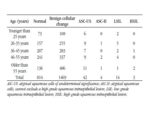
Édina K. Fredrich; Jane D. P. RennerJ. Bras. Patol. Med. Lab. 2019;55(3):246-257DOI: 10.5935/1676-2444.20190023 ABSTRACT OBJECTIVE: To determine the cytopathologic alterations in women who undergo the Papanicolaou exam by the single health system in a laboratory in the city of Santa Cruz do Sul, Rio Grande do Sul, Brazil.METHODOLOGY: Data were collected from cytopathological reports of the year

Cervical cytopathological changes in Pap smear test in the city of Santa Cruz do Sul, Rio Grande do Sul, Brazil Read More »

Evaluation of TP53 Gene Expression in Patients with Childhood Cancer in Northeast Santa Catarina, Brazil

Assessment of cytological and biochemical parameters stability in cerebrospinal fluid

Renan Domingues; Fernando Brunale; Gustavo Bruniera; CarlosJ. Bras. Patol. Med. Lab. 2019;55(3):258-266DOI: 10.5935/1676-2444.20190021 ABSTRACT OBJECTIVE: The aim of this study was to evaluate the stability, in 12 hours, of cytological and biochemical parameters of the cerebrospinal fluid (CSF). Methods: Cerebrospinal fluid (CSF) samples were aliquoted into five different tubes and stored at room temperature. The first

Assessment of cytological and biochemical parameters stability in cerebrospinal fluid Read More »

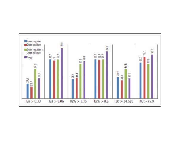
Cut-off values of IG#, IG%, TLC and NC, and capture percentage by group of microorganism IG#: absolute automated count of immature granulocytes; IG%: relative automated count of immature granulocytes; TLC: total leukocyte count; NC: neutrophil count.

Automated immature granulocyte count in patients of the intensive care unit with suspected infection

Letícia R. Lima; Gustavo S. P. Cunha; Keite S. Nogueira; Samuel Ricardo ComarJ. Bras. Patol. Med. Lab. 2019;55(3):267-280DOI: 10.5935/1676-2444.20190031 ABSTRACT INTRODUCTION: Diagnosing infections in intensive care unit (ICU) patients is vital to provide appropriate therapies. Hematological analyzers perform automated immature granulocyte counts (IG) quickly and with no additional cost when compared to traditional microbiological cultures. Elevated

Automated immature granulocyte count in patients of the intensive care unit with suspected infection Read More »

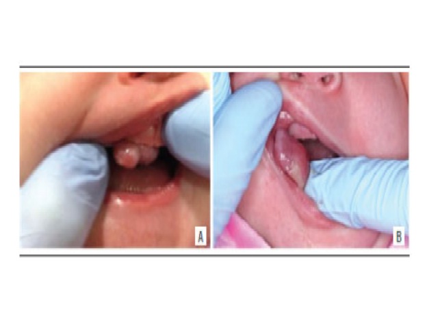
Clinical aspect A and B) clinical aspect of the lesion evidencing a nodule in the alveolar ridge of the anterior maxilla region.

Congenital granular cell epulis: case report and differential diagnosis

Katianne S. Rodrigues; Caio César S. Barros; Ondina Karla M. S. Rocha; Luiz Arthur B. Silva; Marina B. Paies; Márcia Cristina C. MiguelJ. Bras. Patol. Med. Lab. 2019;55(3):281-288DOI: 10.5935/1676-2444.20190026 ABSTRACT A 16-days-old female patient exhibiting an exophytic nodular lesion located at anterior maxilla on the alveolar ridge since birth. The clinical hypothesis of congenital granular

Congenital granular cell epulis: case report and differential diagnosis Read More »

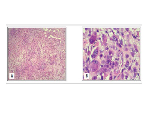
Pathology study with regular HE staining A) low magnification image; B) higher magnification shows markedly proliferative cellular atypia with a myxoid matrix and a high mitotic index. HE: hematoxylin and eosin.

Nodular fasciitis: case report

Hugo Guilherme M. Jurema; Rafael H. Tomaszeski; Lucas R. Mostardeiro; Valéria M. Jorge; Eduardo B. C. BiccaJ. Bras. Patol. Med. Lab. 2019;55(3):281-288DOI: 10.5935/1676-2444.20190028 ABSTRACT Nodular fasciitis (NF) is a rare fibroblast proliferation of unknown etiology, with benign, rapid clonal growth, from a superficial fascia to the subcutaneous tissue or an adjacent muscular layer. Also known

Nodular fasciitis: case report Read More »

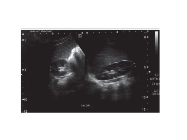
Dynamic US of the right kidney showing kidney size US: ultrasonography.

Case report of staghorn calculi in a pediatric patient

Isadora E. Silva; Gabriela C. Abreu; Wagner M. Moura; Débora C. Cerqueira; Cinthia A. C. LeandroJ. Bras. Patol. Med. Lab. 2019;55(3):295-304DOI: 10.5935/1676-2444.20190022 ABSTRACT This article reports a case of large volume asymptomatic staghorn calculus, and a brief revision of this rare pathology is done. A two-yearold male patient was diagnosed with staghorn calculus and hydronephrosis

Case report of staghorn calculi in a pediatric patient Read More »

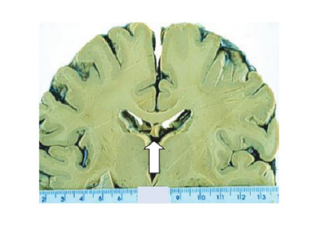
Macroscopic aspect of brain and heart obtained from autopsied patients with cysticercosis, from 1990 to 2018 (arrows)

Autopsies cysticercosis and Aids concomitance

Camila L. Cavellani; Grace Kelly N. A. Favarato; Aline Cristina S. Silva; Lívia F. Oliveira; Ana Carolina G. Faleiros; Rosana Rosa M. Corrêa; Ruy S. Lino-Júnior; Vicente de Paula A. TeixeiraJ. Bras. Patol. Med. Lab. 2019;55(3):310-314DOI: 10.5935/1676-2444.20190025 ABSTRACT The objective of the present study was to report the concomitance of cysticercosis and acquired immunodeficiency syndrome

Autopsies cysticercosis and Aids concomitance Read More »

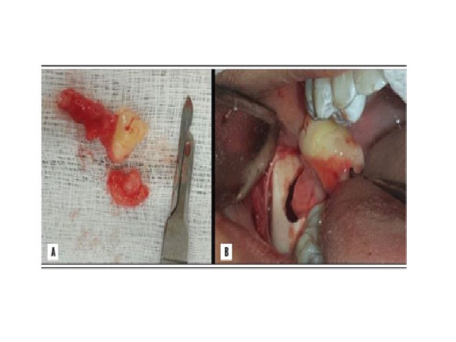
image showing the extension of the tissue associated with the dental element; B) intraoral images showing the moment of tooth 48 removal along with the altered tissue

Hyperplastic dental follicle: case report

Vinicius R. Gomes; Maria Carline S. Melo; Helder C. Carnei Jr.; João Eudes T. Pinho Filho; Murilo A. Teixeira NetoJ. Bras. Patol. Med. Lab. 2019;55(3):315-320DOI: 10.5935/1676-2444.20190027 ABSTRACT The dental follicle is characterized as a radiolucent area around an impacted tooth. It is believed that these follicles can originate cysts and tumors. The aim of this

Hyperplastic dental follicle: case report Read More »

Evaluation of TP53 Gene Expression in Patients with Childhood Cancer in Northeast Santa Catarina, Brazil

The unusual paradox of cancer-associated inflammation: an update

Cláudia Roberta L. V. FigueiredoJ. Bras. Patol. Med. Lab. 2019;55(3):321-332DOI: 10.5935/1676-2444.20190029 ABSTRACT The inflammatory response represents a fundamental component of the tumor microenvironment and is responsible for mediating the biological communication network and the molecular signal flow that characterize the neoplastic tissue. Thus, influenced by the inflammatory process, neoplastic and non-neoplastic cells (recruited stromal and

The unusual paradox of cancer-associated inflammation: an update Read More »