Brazilian Journal of Pathology and Laboratory Medicine

Please contact: journal@jbpml.org.br

Pathology

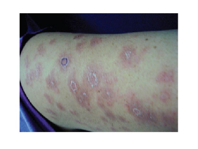
Clinical aspect of patient with metastatic cutaneous Crohn's disease: erythematous scaly plates in left arm

Metastatic cutaneous Crohn’s disease as an important differential diagnosis of granulomatous skin disease

Murilo C. Peretti; Andressa T. Szczypkovski; Gabriel S. Reis; Gabriela M. Quadros; Maira M. Mukai; Heda Maria B. S. Amarante; Betina WernerJ. Bras. Patol. Med. Lab. 2016;52(2):112-11510.5935/1676-2444.20160013 ABSTRACT Patients with Crohn’s disease may show extraintestinal manifestations, including cutaneous, whose frequency ranges from 2% to 34%. Metastatic cutaneous Crohn’s disease is considered a specific and rare […]

Metastatic cutaneous Crohn’s disease as an important differential diagnosis of granulomatous skin disease Read More »

Underreporting of scientific knowledge: a theme for reflection

Report of two cases of vacuolar interface dermatitis initially suspected as melanoma in situ and review of the literature

Relato de dois casos de dermatite de interface vacuolar, inicialmente diagnosticados como melanoma in situ, e revisão da literatura Luciana Pantaleão1; Rosane Orofino-Costa2; Mariléia C. Torres3; Rossitza Lazova4; Mayra C. Rochael1 1. Universidade Federal Fluminense (UFF), Rio de Janeiro, Brasil2. Universidade do Estado do Rio de Janeiro (UERJ), Rio de Janeiro, Brasil3. Diagnose Anatomia Patológica e

Report of two cases of vacuolar interface dermatitis initially suspected as melanoma in situ and review of the literature Read More »

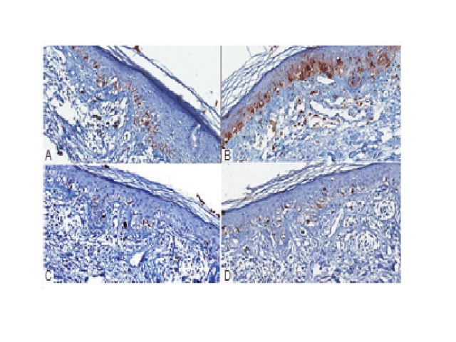
A) numerous isolated cells in the basal layer, in a continuous pattern, labeled with Melan-A, mimicking melanoma in situ; B) S100 stain highlighted junctional melanocytes as well as langerhans cells in the epidermis; C and D) a normal number of melanocytes labeled with HMB45 (C) and tyrosinase (D)

Report of two cases of vacuolar interface dermatitis initially suspected as melanoma in situ and review of the literature

Luciana Pantaleão; Rosane Orofino-Costa; Mariléia C. Torres; Rossitza Lazova; Mayra C. RochaelJ. Bras. Patol. Med. Lab. 2016;52(2):116-119DOI: 10.5935/1676-2444.20160015 ABSTRACT The diagnosis of melanocytic lesions can be challenging and immunohistochemical study is a valuable tool for dermatopathologists. We report two cases initially simulating melanoma in situ, reviewing the histopathological and immunohistochemical findings and the cases published in

Report of two cases of vacuolar interface dermatitis initially suspected as melanoma in situ and review of the literature Read More »

Underreporting of scientific knowledge: a theme for reflection

Proliferating trichilemmal tumor: case report

Tumor triquilemal proliferante: relato de caso Theresinha C. Fonseca; Camila L. Bandeira; Bruno A. Sousa; Terence P. Farias; Ana Lúcia A. Eisenberg Instituto Nacional de Câncer (Inca) José Alencar Gomes da Silva, Rio de Janeiro, Brasil DOI: 10.5935/1676-2444.20160014 Corresponding author Theresinha Carvalho da FonsecaInstituto Nacional de CâncerAv. Cordeiro da Graça, 156; Santo CristoCEP: 20220-400; Rio

Proliferating trichilemmal tumor: case report Read More »

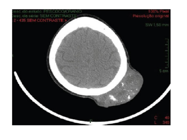
Computed tomography, axial view Hypodense scalp lesion, with calcifications without periosteal invasion, in left parietaltemporal region.

Proliferating trichilemmal tumor: case report

Theresinha C. Fonseca; Camila L. Bandeira; Bruno A. Sousa; Terence P. Farias; Ana Lúcia A. EisenbergJ. Bras. Patol. Med. Lab. 2016;52(2):120-123DOI: 10.5935/1676-2444.20160014 ABSTRACT The proliferating trichilemmal tumor is a lesion with trichilemmal differentiation, more common occurring among elderly women; it affects the scalp with sizes ranging from 2-10 cm. Microscopically, it is solid-cystic, well-defined, affecting

Proliferating trichilemmal tumor: case report Read More »

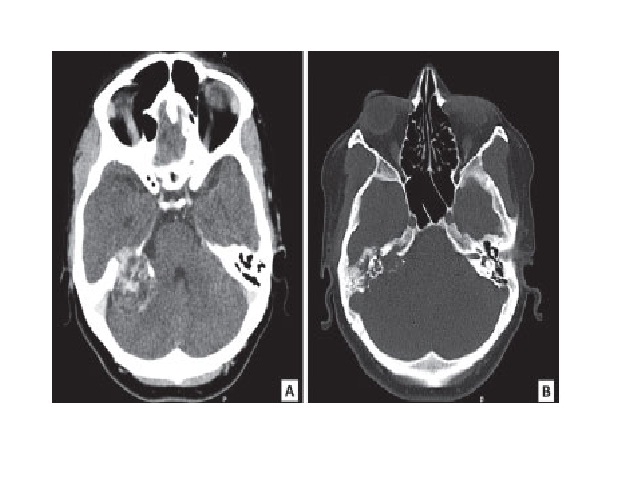
Aggressive papillary tumor of endolymphatic sac: CT showing an expansile lytic lesion arising in the right temporal bone (A and B) CT: computed tomography.

Aggressive papillary tumor of endolymphatic sac: case report of a rare neoplasia

Eduardo Cambruzzi; Karla Lais Pêgas; Leandro P. Almeida; Gerson Evandro Perondi; Leandro I. DiniJ. Bras. Patol. Med. Lab. 2016;52(1):31-34DOI: 10.5935/1676-2444.20160010 ABSTRACT Aggressive papillary endolymphatic sac tumor (ELST) is a rare neoplasm, occasionally related to von Hippel-Lindau disease, characterized by locally aggressive growth with temporal bone destruction. The authors report a case of ELST in a

Aggressive papillary tumor of endolymphatic sac: case report of a rare neoplasia Read More »

Underreporting of scientific knowledge: a theme for reflection

Aggressive papillary tumor of endolymphatic sac: case report of a rare neoplasia

Tumor papilar agressivo do saco endolinfático: relato de caso de uma rara neoplasia Eduardo Cambruzzi1; Karla Lais Pêgas2; Leandro P. Almeida3; Gerson Evandro Perondi3; Leandro I. Dini3 1. Instituto de Cardiologia – Fundação Universitária de Cardiologia (ICFUC), Rio Grande do Sul, Brasil2. Santa Casa de Porto Alegre, Rio Grande do Sul, Brasil3. Hospital Cristo Redentor,

Aggressive papillary tumor of endolymphatic sac: case report of a rare neoplasia Read More »

Underreporting of scientific knowledge: a theme for reflection

Aggressive angiomyxoma of the vagina: a case report

Angiomixoma agressivo de vagina: um relato de caso Matheo Augusto M. Stumpf; Rebecca S. M. Stival; Alexandre B. Merlini; Fábio P. Mansani; Janicelli B. C. H. Silvestre; Isabela B. Mongruel; Mário R. Montemór Netto Universidade Estadual de Ponta Grossa (UEPG), Paraná, Brasil DOI: 10.5935/1676-2444.20160011 Corresponding author Mário Rodrigues Montemór NettoRua Santos Dumont, 1.436; CentroCEP: 84010-360;

Aggressive angiomyxoma of the vagina: a case report Read More »

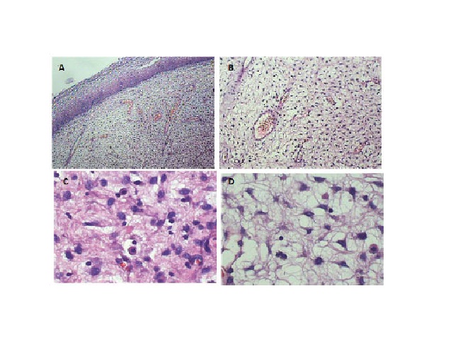
AAM in HE staining revealing a fragment of squamous mucosa with spindlecell neoplastic proliferation in loose collagenous stroma with a myxoid matrix embedding irregular vessels and inflammatory cells. A) 40×; B) 100×; C) 400×; D) 400× AAM: aggressive angiomyxoma; HE: hematoxylin and eosin.

Aggressive angiomyxoma of the vagina: a case report

Matheo Augusto M. Stumpf; Rebecca S. M. Stival; Alexandre B. Merlini; Fábio P. Mansani; Janicelli B. C. H. Silvestre; Isabela B. Mongruel; Mário R. Montemór NettoJ. Bras. Patol. Med. Lab. 2016;52(1):35-38DOI: 10.5935/1676-2444.20160011 ABSTRACT Aggressive angiomyxoma (AAM) is a rare infiltrative tumor of mesenchymal origin that has high rates of local recurrence. We present the case

Aggressive angiomyxoma of the vagina: a case report Read More »

Underreporting of scientific knowledge: a theme for reflection

Elastosis perforans serpiginosa, a transepithelial elimination skin disease diagnosed by histopathology: case report

Elastose perfurante serpiginosa, uma doença de transeliminação epidérmica e diagnóstico essencialmente histopatológico: relato de caso Alexandre M. Lima1; Milena M. Rodrigues1; Sheila P. Rocha2; Cibele C. Rodrigues2; Carmelia M. S. Reis2 1. Universidade Federal de Mato Grosso do Sul (UFMS), Mato Grosso do Sul, Brasil2. Hospital Regional da Asa Norte (HRAN), Brasília, Brasil DOI: 10.5935/1676-2444.20160004

Elastosis perforans serpiginosa, a transepithelial elimination skin disease diagnosed by histopathology: case report Read More »