Gingival carcinoma cuniculatum mimicking a reactive/inflammatory process
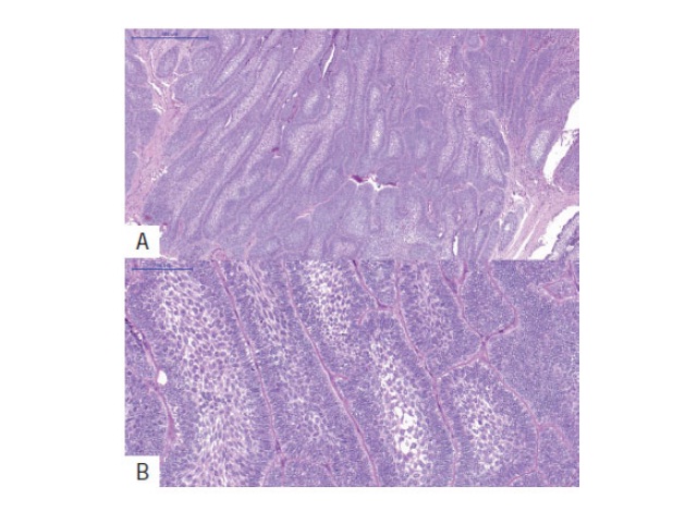
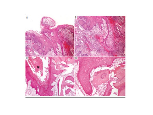
Rodrigo N. Silva, Heitor A. Silveira, Luciana Y. Almeida, Carla N. B. Colturato, Alexandre Elias Trivellato, Cassio E. Sverzut, Jorge E. LeónJ. Bras. Patol. Med. Lab. 2019;55(5):498-505DOI: 10.5935/1676-2444.20190045 ABSTRACT Carcinoma cuniculatum (CC), a rare variant of oral squamous cell carcinoma, presents well-differentiated neoplastic epithelial cells infiltrating the underlying submucosal or bone tissues, forming the so-called […]
Gingival carcinoma cuniculatum mimicking a reactive/inflammatory process Read More »