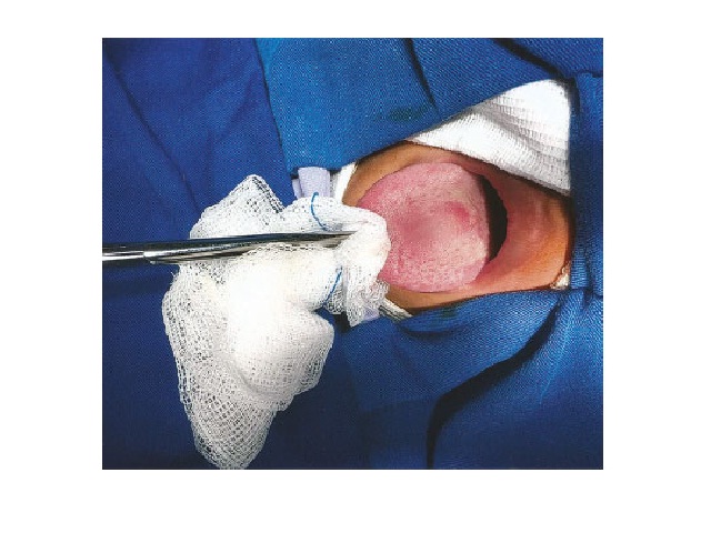
Peripheral odontogenic fibroma in the mandibular gingiva: case report
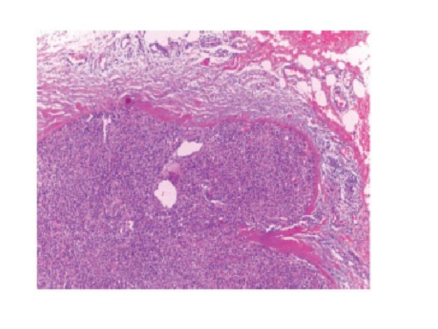
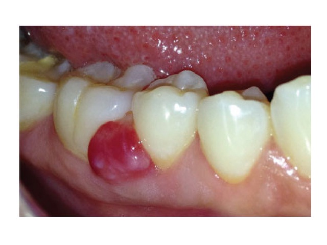
Luana Samara B. de-Sena; Márcia Cristina C. Miguel; Jozinete V. Pereira; Daliana Q. C. Gomes; Pollianna M. Alves; Cassiano Francisco W. NonakaJ. Bras. Patol. Med. Lab. 2019;55(2):192-201DOI: 10.5935/1676-2444.20190015 ABSTRACT Peripheral odontogenic fibroma (POF) is a rare benign neoplasm of odontogenic mesenchymal origin that accounts for approximately 4.7% of all odontogenic tumors. This article reports the […]
Peripheral odontogenic fibroma in the mandibular gingiva: case report Read More »