Brazilian Journal of Pathology and Laboratory Medicine

Please contact: journal@jbpml.org.br

Case Report

Underreporting of scientific knowledge: a theme for reflection

Giant gastrointestinal stromal tumor of the proximal ileum

Tumor estromal gastrointestinal gigante do íleo proximal Kelen Christina A. Bezzerra Hospital São Francisco, Concórdia, Santa Catarina, Brazil Corresponding author Kelen Christina Alves BezzerraORCID 0000-0001-7914-432Xe-mail: drpatologia@hospitalsaofrancisco.com First Submission on 3/29/2019 Last Submission on 4/3/2019Accepted for publication on 4/3/2019Published on 8/20/2019 ABSTRACT The gastrointestinal stromal tumors (GIST) are rare and consist in mesenchymal neoplasms of the gastrointestinal tract, which may affect […]

Giant gastrointestinal stromal tumor of the proximal ileum Read More »

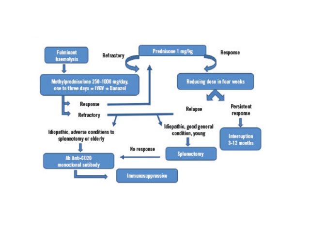
Flowchart of warm AHAI treatment AIHA: autoimmune haemolytic anemia; IVGV: intravenous immunoglobulins; Ab: antibody.

Evans syndrome in male lupus patient: case report

Barbara F. Dalmaso, Afonso José P. Cortez, Maitê Alexandra E. Velasquez, Warlindo C. Silva Neto, Flávio Aparecido R. Silva, Lívia Mara S. SousaJ. Bras. Patol. Med. Lab. 2019;55(4):416-425DOI: 10.5935/1676-2444.20190038 ABSTRACT Evans syndrome (ES) is a rare autoimmune disease of unknown etiology. ES occurs when there is a combination of autoimmune hemolytic anemia (AIHA) with thrombocytopenia,

Evans syndrome in male lupus patient: case report Read More »

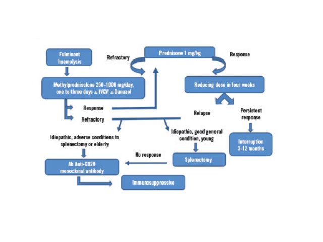
Flowchart of warm AHAI treatment AIHA: autoimmune haemolytic anemia; IVGV: intravenous immunoglobulins; Ab: antibody.

Evans syndrome in male lupus patient: case report

Síndrome de Evans em paciente lúpico do sexo masculino: relato de caso Barbara F. Dalmaso, Afonso José P. Cortez, Maitê Alexandra E. Velasquez, Warlindo C. Silva Neto, Flávio Aparecido R. Silva, Lívia Mara S. Sousa Universidade Santo Amaro São Paulo, São Paulo, Brazil Corresponding author Barbara Ferraço DalmasoORCID 0000-0003-1311-3496e-mail: babi_dalmaso@hotmail.com First Submission on 3/28/2019 Last Submission on 4/3/2019Accepted

Evans syndrome in male lupus patient: case report Read More »

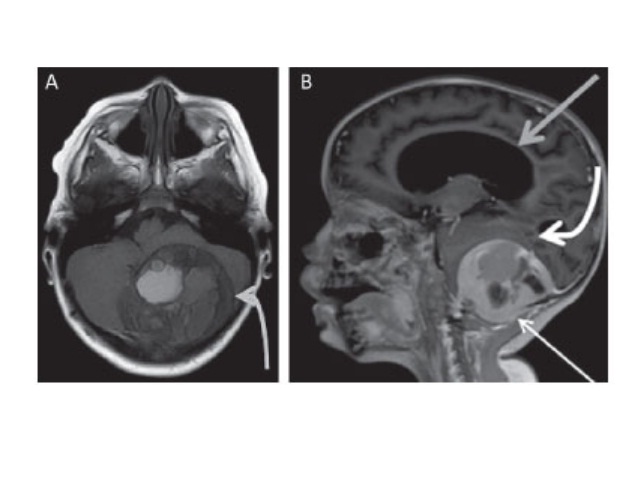
A) MRI in T1-weighted axial image without contrast demonstrating well-defined extra-axial mass in the posterior fossa (curved light gray arrow), projecting to the adjacent soft parts; B) MRI, T1-weighted post-contrast sagittal image demonstrating well-defined extra-axial mass in the posterior fossa traversing the posterolateral inferior occipital bone, projecting to the adjacent soft parts, with intense peripheral enhancement by contrast (fine white arrow), striking the cerebellar hemispheres (curved arrow) and distorting the fourth ventricle with severe hydrocephalus upstream (gray arrow) MRI: magnetic resonance imaging.

Aggressive osteoblastoma with degeneration to secondary aneurysmal bone cyst

Marcio Luís Duarte; José Luiz M. A. Prado; Carlos Alexandre M. Pereira; Maria Teresa S. Alves; Luiz Carlos D. ScoppettaJ. Bras. Patol. Med. Lab. 2019;55(4):426-433DOI: 10.5935/1676-2444.20190039 ABSTRACT A 4-year-old male patient presented with headaches that started 18 months ago and were associated with progressive loss of balance and difficulty walking. Magnetic resonance imaging (MRI) demonstrated

Aggressive osteoblastoma with degeneration to secondary aneurysmal bone cyst Read More »

Underreporting of scientific knowledge: a theme for reflection

Aggressive osteoblastoma with degeneration to secondary aneurysmal bone cyst

Osteoblastoma agressivo com degeneração secundária para cisto ósseo aneurismático Marcio Luís Duarte1; José Luiz M. A. Prado1; Carlos Alexandre M. Pereira2; Maria Teresa S. Alves3; Luiz Carlos D. Scoppetta2 1. Webimagem, São Paulo, São Paulo, Brazil2. Hospital São Camilo, São Paulo, São Paulo, Brazil3. Universidade Federal de São Paulo (Unifesp), São Paulo, São Paulo, Brazil

Aggressive osteoblastoma with degeneration to secondary aneurysmal bone cyst Read More »

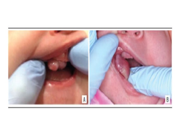
Clinical aspect A and B) clinical aspect of the lesion evidencing a nodule in the alveolar ridge of the anterior maxilla region.

Congenital granular cell epulis: case report and differential diagnosis

Katianne S. Rodrigues; Caio César S. Barros; Ondina Karla M. S. Rocha; Luiz Arthur B. Silva; Marina B. Paies; Márcia Cristina C. MiguelJ. Bras. Patol. Med. Lab. 2019;55(3):281-288DOI: 10.5935/1676-2444.20190026 ABSTRACT A 16-days-old female patient exhibiting an exophytic nodular lesion located at anterior maxilla on the alveolar ridge since birth. The clinical hypothesis of congenital granular

Congenital granular cell epulis: case report and differential diagnosis Read More »

Evaluation of TP53 Gene Expression in Patients with Childhood Cancer in Northeast Santa Catarina, Brazil

Congenital granular cell epulis: case report and differential diagnosis

Epúlide de células granulares congênita: relato de caso e diagnóstico diferencial Katianne S. Rodrigues; Caio César S. Barros; Ondina Karla M. S. Rocha; Luiz Arthur B. Silva; Marina B. Paies; Márcia Cristina C. Miguel Epúlide de células granulares congênita: relato de caso e diagnóstico diferencial Corresponding author Márcia Cristina da Costa MiguelORCID: 0000-0002-6661-2566e-mail: mccmiguel@hotmail.com First Submission

Congenital granular cell epulis: case report and differential diagnosis Read More »

Underreporting of scientific knowledge: a theme for reflection

Nodular fasciitis: case report

Fasciíte nodular: relato de caso Hugo Guilherme M. Jurema1; Rafael H. Tomaszeski1; Lucas R. Mostardeiro1; Valéria M. Jorge2; Eduardo B. C. Bicca1 1. Universidade Católica de Pelotas (UCPel), Pelotas, Rio Grande do Sul, Brazil2. Universidade Federal de Pelotas (UFPel), Pelotas, Rio Grande do Sul, Brazil Corresponding author Hugo Guilherme de Moraes JuremaORCID: 0000-0003-4692-0470e-mail: hugogmj@gmail.com First Submission

Nodular fasciitis: case report Read More »

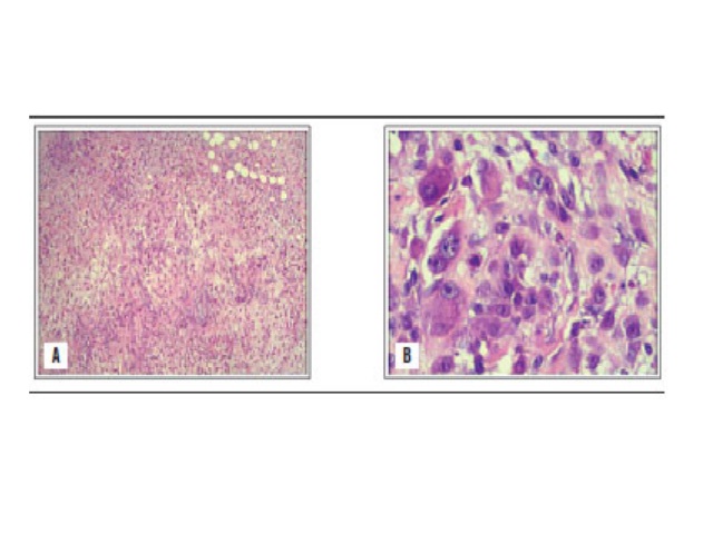
Pathology study with regular HE staining A) low magnification image; B) higher magnification shows markedly proliferative cellular atypia with a myxoid matrix and a high mitotic index. HE: hematoxylin and eosin.

Nodular fasciitis: case report

Hugo Guilherme M. Jurema; Rafael H. Tomaszeski; Lucas R. Mostardeiro; Valéria M. Jorge; Eduardo B. C. BiccaJ. Bras. Patol. Med. Lab. 2019;55(3):281-288DOI: 10.5935/1676-2444.20190028 ABSTRACT Nodular fasciitis (NF) is a rare fibroblast proliferation of unknown etiology, with benign, rapid clonal growth, from a superficial fascia to the subcutaneous tissue or an adjacent muscular layer. Also known

Nodular fasciitis: case report Read More »

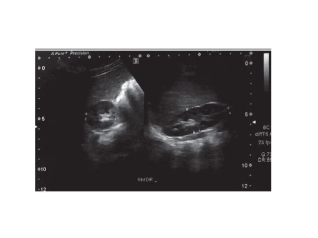
Dynamic US of the right kidney showing kidney size US: ultrasonography.

Case report of staghorn calculi in a pediatric patient

Isadora E. Silva; Gabriela C. Abreu; Wagner M. Moura; Débora C. Cerqueira; Cinthia A. C. LeandroJ. Bras. Patol. Med. Lab. 2019;55(3):295-304DOI: 10.5935/1676-2444.20190022 ABSTRACT This article reports a case of large volume asymptomatic staghorn calculus, and a brief revision of this rare pathology is done. A two-yearold male patient was diagnosed with staghorn calculus and hydronephrosis

Case report of staghorn calculi in a pediatric patient Read More »