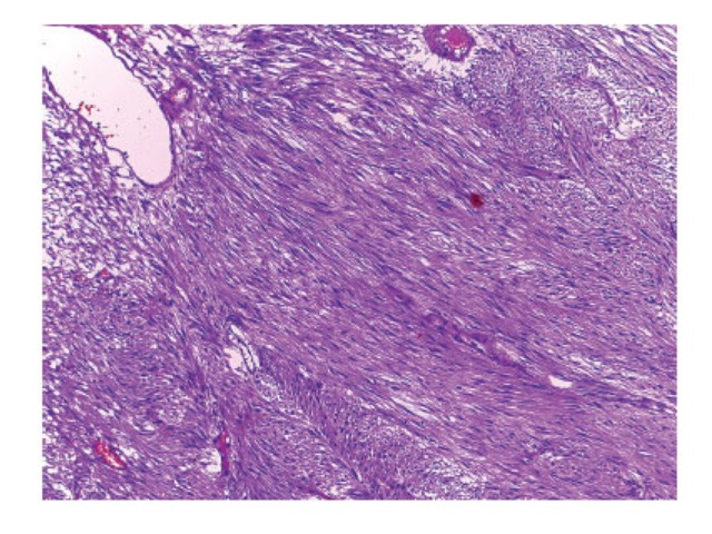
Dermatoscopy in polypoid basal-cell carcinoma: a rare histopathological variation
Dermatoscopia em carcinoma basocelular polipoide: uma variante histopatológica rara Mariana A. Almeida, Gabriella C. Carmo, Michele R. Feroldi, Gustavo Verardino Universidade de Taubaté, Taubaté, São Paulo, Brazil2. Instituto Nacional do Câncer (Inca), Rio de Janeiro, Rio de Janeiro, Brazil3. Clínica Dér Médical, Campos do Jordão, São Paulo, Brazil DOI: 10.5935/1676-2444.20190047 Corresponding author Mariana Abdo de […]
Dermatoscopy in polypoid basal-cell carcinoma: a rare histopathological variation Read More »