Brazilian Journal of Pathology and Laboratory Medicine

Please contact: journal@jbpml.org.br

cadmin

Cervical cytopathological changes in Pap smear test in the city of Santa Cruz do Sul, Rio Grande do Sul, Brazil

Cervical cytopathological changes in Pap smear test in the city of Santa Cruz do Sul, Rio Grande do Sul, Brazil

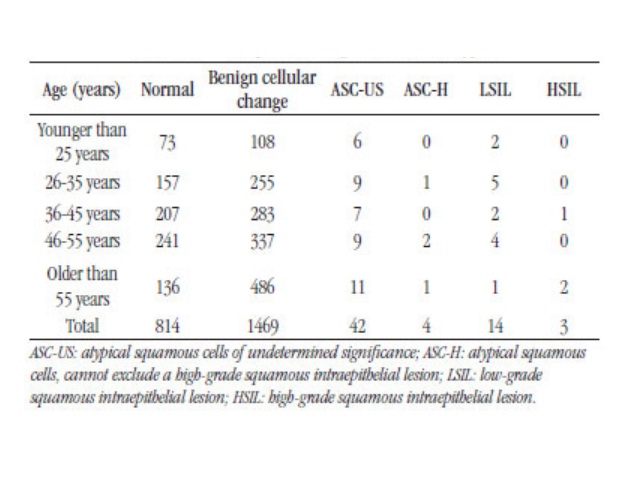
Édina K. Fredrich; Jane D. P. RennerJ. Bras. Patol. Med. Lab. 2019;55(3):246-257DOI: 10.5935/1676-2444.20190023 ABSTRACT OBJECTIVE: To determine the cytopathologic alterations in women who undergo the Papanicolaou exam by the single health system in a laboratory in the city of Santa Cruz do Sul, Rio Grande do Sul, Brazil.METHODOLOGY: Data were collected from cytopathological reports of the year […]

Cervical cytopathological changes in Pap smear test in the city of Santa Cruz do Sul, Rio Grande do Sul, Brazil Read More »

Underreporting of scientific knowledge: a theme for reflection

Cervical cytopathological changes in Pap smear test in the city of Santa Cruz do Sul, Rio Grande do Sul, Brazil

Alterações citopatológicas em exames de Papanicolaou na cidade de Santa Cruz do Sul, Rio Grande do Sul, Brasil Édina K. Fredrich; Jane D. P. Renner Universidade de Santa Cruz do Sul (UNISC), Santa Cruz do Sul, Rio Grande do Sul, Brazil Corresponding author Édina Knod FredrichOrcid: 0000-0002-5585-0649e-mail: ediina.02@gmail.com First Submission on 9/6/2018 Last Submission on 12/5/2018Accepted for publication

Cervical cytopathological changes in Pap smear test in the city of Santa Cruz do Sul, Rio Grande do Sul, Brazil Read More »

Evaluation of TP53 Gene Expression in Patients with Childhood Cancer in Northeast Santa Catarina, Brazil

Assessment of cytological and biochemical parameters stability in cerebrospinal fluid

Renan Domingues; Fernando Brunale; Gustavo Bruniera; CarlosJ. Bras. Patol. Med. Lab. 2019;55(3):258-266DOI: 10.5935/1676-2444.20190021 ABSTRACT OBJECTIVE: The aim of this study was to evaluate the stability, in 12 hours, of cytological and biochemical parameters of the cerebrospinal fluid (CSF). Methods: Cerebrospinal fluid (CSF) samples were aliquoted into five different tubes and stored at room temperature. The first

Assessment of cytological and biochemical parameters stability in cerebrospinal fluid Read More »

Underreporting of scientific knowledge: a theme for reflection

Assessment of cytological and biochemical parameters stability in cerebrospinal fluid

Renan Domingues; Fernando Brunale; Gustavo Bruniera; Carlos Senne Liquor Diagnóstico, São Paulo, São Paulo, Brazil Corresponding author Renan DominguesORCID: 0000-0002-6058-7937e-mail: renan.domingues@senneliquor.com.br First Submission on 7/24/2018 Last Submission on 7/25/2018Accepted for publication on 3/19/2019Published on 6/20/2019 ABSTRACT OBJECTIVE: The aim of this study was to evaluate the stability, in 12 hours, of cytological and biochemical parameters of the cerebrospinal fluid (CSF). Methods:

Assessment of cytological and biochemical parameters stability in cerebrospinal fluid Read More »

Underreporting of scientific knowledge: a theme for reflection

Automated immature granulocyte count in patients of the intensive care unit with suspected infection

Contagem automatizada de granulócitos imaturos em pacientes de uma unidade de terapia intensiva com suspeita de infecção Letícia R. Lima1; Gustavo S. P. Cunha2; Keite S. Nogueira1; Samuel Ricardo Comar1 1. Complexo Hospital de Clínicas da Universidade Federal do Paraná (CHC-UFPR); Curitiba, Paraná, Brasil2. Universidade Federal do Paraná (UFPR); Curitiba, Paraná, Brasil Corresponding author Samuel

Automated immature granulocyte count in patients of the intensive care unit with suspected infection Read More »

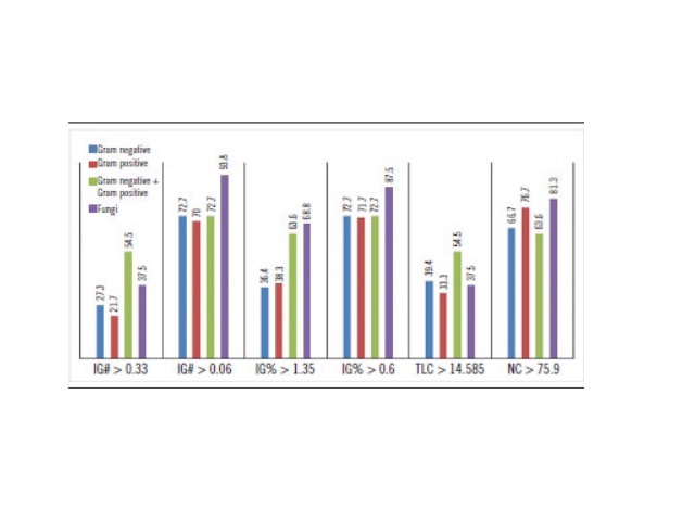
Cut-off values of IG#, IG%, TLC and NC, and capture percentage by group of microorganism IG#: absolute automated count of immature granulocytes; IG%: relative automated count of immature granulocytes; TLC: total leukocyte count; NC: neutrophil count.

Automated immature granulocyte count in patients of the intensive care unit with suspected infection

Letícia R. Lima; Gustavo S. P. Cunha; Keite S. Nogueira; Samuel Ricardo ComarJ. Bras. Patol. Med. Lab. 2019;55(3):267-280DOI: 10.5935/1676-2444.20190031 ABSTRACT INTRODUCTION: Diagnosing infections in intensive care unit (ICU) patients is vital to provide appropriate therapies. Hematological analyzers perform automated immature granulocyte counts (IG) quickly and with no additional cost when compared to traditional microbiological cultures. Elevated

Automated immature granulocyte count in patients of the intensive care unit with suspected infection Read More »

Evaluation of TP53 Gene Expression in Patients with Childhood Cancer in Northeast Santa Catarina, Brazil

Congenital granular cell epulis: case report and differential diagnosis

Epúlide de células granulares congênita: relato de caso e diagnóstico diferencial Katianne S. Rodrigues; Caio César S. Barros; Ondina Karla M. S. Rocha; Luiz Arthur B. Silva; Marina B. Paies; Márcia Cristina C. Miguel Epúlide de células granulares congênita: relato de caso e diagnóstico diferencial Corresponding author Márcia Cristina da Costa MiguelORCID: 0000-0002-6661-2566e-mail: mccmiguel@hotmail.com First Submission

Congenital granular cell epulis: case report and differential diagnosis Read More »

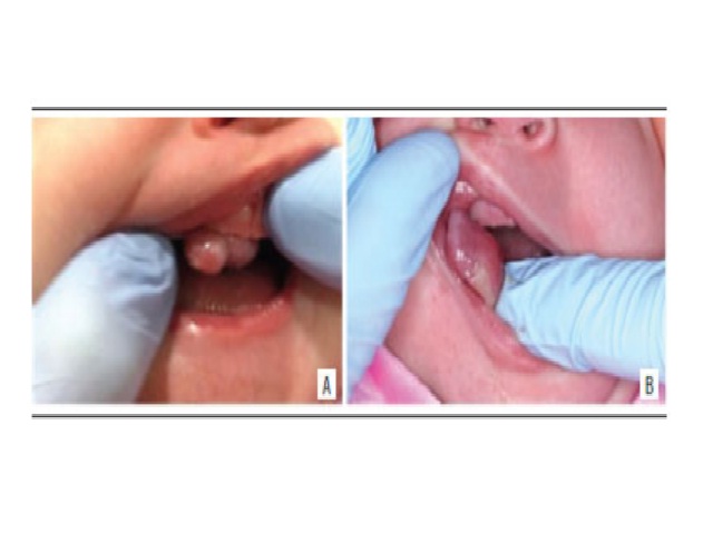
Clinical aspect A and B) clinical aspect of the lesion evidencing a nodule in the alveolar ridge of the anterior maxilla region.

Congenital granular cell epulis: case report and differential diagnosis

Katianne S. Rodrigues; Caio César S. Barros; Ondina Karla M. S. Rocha; Luiz Arthur B. Silva; Marina B. Paies; Márcia Cristina C. MiguelJ. Bras. Patol. Med. Lab. 2019;55(3):281-288DOI: 10.5935/1676-2444.20190026 ABSTRACT A 16-days-old female patient exhibiting an exophytic nodular lesion located at anterior maxilla on the alveolar ridge since birth. The clinical hypothesis of congenital granular

Congenital granular cell epulis: case report and differential diagnosis Read More »

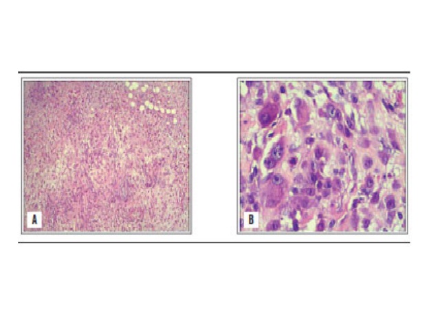
Pathology study with regular HE staining A) low magnification image; B) higher magnification shows markedly proliferative cellular atypia with a myxoid matrix and a high mitotic index. HE: hematoxylin and eosin.

Nodular fasciitis: case report

Hugo Guilherme M. Jurema; Rafael H. Tomaszeski; Lucas R. Mostardeiro; Valéria M. Jorge; Eduardo B. C. BiccaJ. Bras. Patol. Med. Lab. 2019;55(3):281-288DOI: 10.5935/1676-2444.20190028 ABSTRACT Nodular fasciitis (NF) is a rare fibroblast proliferation of unknown etiology, with benign, rapid clonal growth, from a superficial fascia to the subcutaneous tissue or an adjacent muscular layer. Also known

Nodular fasciitis: case report Read More »

Underreporting of scientific knowledge: a theme for reflection

Nodular fasciitis: case report

Fasciíte nodular: relato de caso Hugo Guilherme M. Jurema1; Rafael H. Tomaszeski1; Lucas R. Mostardeiro1; Valéria M. Jorge2; Eduardo B. C. Bicca1 1. Universidade Católica de Pelotas (UCPel), Pelotas, Rio Grande do Sul, Brazil2. Universidade Federal de Pelotas (UFPel), Pelotas, Rio Grande do Sul, Brazil Corresponding author Hugo Guilherme de Moraes JuremaORCID: 0000-0003-4692-0470e-mail: hugogmj@gmail.com First Submission

Nodular fasciitis: case report Read More »