Brazilian Journal of Pathology and Laboratory Medicine

Please contact: journal@jbpml.org.br

cadmin

Aggressive papillary tumor of endolymphatic sac: CT showing an expansile lytic lesion arising in the right temporal bone (A and B) CT: computed tomography.

Aggressive papillary tumor of endolymphatic sac: case report of a rare neoplasia

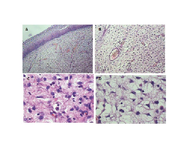
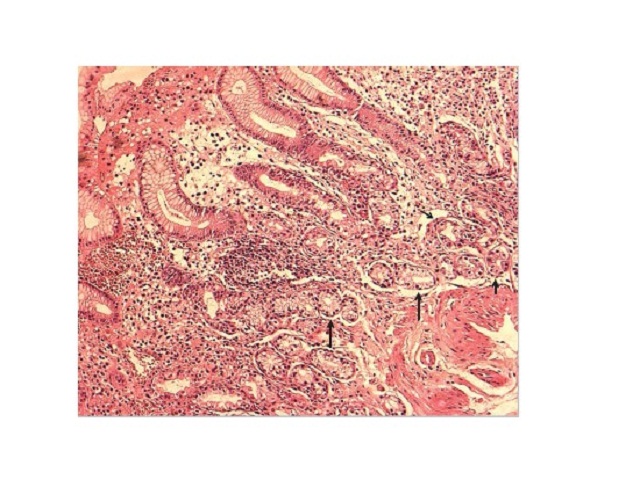
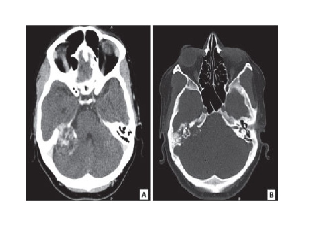
Eduardo Cambruzzi; Karla Lais Pêgas; Leandro P. Almeida; Gerson Evandro Perondi; Leandro I. DiniJ. Bras. Patol. Med. Lab. 2016;52(1):31-34DOI: 10.5935/1676-2444.20160010 ABSTRACT Aggressive papillary endolymphatic sac tumor (ELST) is a rare neoplasm, occasionally related to von Hippel-Lindau disease, characterized by locally aggressive growth with temporal bone destruction. The authors report a case of ELST in a […]

Aggressive papillary tumor of endolymphatic sac: case report of a rare neoplasia Read More »

Underreporting of scientific knowledge: a theme for reflection

Aggressive papillary tumor of endolymphatic sac: case report of a rare neoplasia

Tumor papilar agressivo do saco endolinfático: relato de caso de uma rara neoplasia Eduardo Cambruzzi1; Karla Lais Pêgas2; Leandro P. Almeida3; Gerson Evandro Perondi3; Leandro I. Dini3 1. Instituto de Cardiologia – Fundação Universitária de Cardiologia (ICFUC), Rio Grande do Sul, Brasil2. Santa Casa de Porto Alegre, Rio Grande do Sul, Brasil3. Hospital Cristo Redentor,

Aggressive papillary tumor of endolymphatic sac: case report of a rare neoplasia Read More »

Underreporting of scientific knowledge: a theme for reflection

Aggressive angiomyxoma of the vagina: a case report

Angiomixoma agressivo de vagina: um relato de caso Matheo Augusto M. Stumpf; Rebecca S. M. Stival; Alexandre B. Merlini; Fábio P. Mansani; Janicelli B. C. H. Silvestre; Isabela B. Mongruel; Mário R. Montemór Netto Universidade Estadual de Ponta Grossa (UEPG), Paraná, Brasil DOI: 10.5935/1676-2444.20160011 Corresponding author Mário Rodrigues Montemór NettoRua Santos Dumont, 1.436; CentroCEP: 84010-360;

Aggressive angiomyxoma of the vagina: a case report Read More »

AAM in HE staining revealing a fragment of squamous mucosa with spindlecell neoplastic proliferation in loose collagenous stroma with a myxoid matrix embedding irregular vessels and inflammatory cells. A) 40×; B) 100×; C) 400×; D) 400× AAM: aggressive angiomyxoma; HE: hematoxylin and eosin.

Aggressive angiomyxoma of the vagina: a case report

Matheo Augusto M. Stumpf; Rebecca S. M. Stival; Alexandre B. Merlini; Fábio P. Mansani; Janicelli B. C. H. Silvestre; Isabela B. Mongruel; Mário R. Montemór NettoJ. Bras. Patol. Med. Lab. 2016;52(1):35-38DOI: 10.5935/1676-2444.20160011 ABSTRACT Aggressive angiomyxoma (AAM) is a rare infiltrative tumor of mesenchymal origin that has high rates of local recurrence. We present the case

Aggressive angiomyxoma of the vagina: a case report Read More »

Erythematous keratotic papules arranged in a semi-circular pattern in the neck

Elastosis perforans serpiginosa, a transepithelial elimination skin disease diagnosed by histopathology: case report

Alexandre M. Lima; Milena M. Rodrigues; Sheila P. Rocha; Cibele C. Rodrigues; Carmelia M. S. ReisJ. Bras. Patol. Med. Lab. 2016;52(1):39-42DOI: 10.5935/1676-2444.20160004 ABSTRACT Elastosis perforans serpiginosa is a disease that belongs to the group of perforating dermatoses, in which dermal elastic tissue extrusion occurs through epidermis. It generally affects young male individuals, and is clinically

Elastosis perforans serpiginosa, a transepithelial elimination skin disease diagnosed by histopathology: case report Read More »

Underreporting of scientific knowledge: a theme for reflection

Elastosis perforans serpiginosa, a transepithelial elimination skin disease diagnosed by histopathology: case report

Elastose perfurante serpiginosa, uma doença de transeliminação epidérmica e diagnóstico essencialmente histopatológico: relato de caso Alexandre M. Lima1; Milena M. Rodrigues1; Sheila P. Rocha2; Cibele C. Rodrigues2; Carmelia M. S. Reis2 1. Universidade Federal de Mato Grosso do Sul (UFMS), Mato Grosso do Sul, Brasil2. Hospital Regional da Asa Norte (HRAN), Brasília, Brasil DOI: 10.5935/1676-2444.20160004

Elastosis perforans serpiginosa, a transepithelial elimination skin disease diagnosed by histopathology: case report Read More »

Underreporting of scientific knowledge: a theme for reflection

HPV detection in floor of mouth squamous cell carcinoma by PCR amplification

Detecção do HPV em carcinoma espinocelular de assoalho de boca por amplificação da PCR Luciana E. Simonato1; Saygo Tomo1; José Fernando Garcia2; Luiz Alberto Veronese2; Glauco I. Miyahara2 1. Universidade Camilo Castelo Branco (Unicastelo), São Paulo, Brasil2. Universidade Estadual Paulista Júlio de Mesquita Filho (Unesp), São Paulo, Brasil DOI: 10.5935/1676-2444.20160005 Corresponding author Saygo TomoRua Laudemiro

HPV detection in floor of mouth squamous cell carcinoma by PCR amplification Read More »

Electrophoresis in 8% polyacrylamide gel stained with silver nitrate. HPV amplification (142 bp) HPV: human papillomavirus; MW: 100 bp DNA ladder; HeLa: DNA from HPV 18-infected cervical carcinoma cell line; CO2: condyloma DNA; NO: no DNA control; DNA: deoxyribonucleic acid; MW: molecular weight.

HPV detection in floor of mouth squamous cell carcinoma by PCR amplification

Luciana E. Simonato; Saygo Tomo; José Fernando Garcia; Luiz Alberto Veronese; Glauco I. MiyaharaJ. Bras. Patol. Med. Lab. 2016;52(1):43-49DOI: 10.5935/1676-2444.20160005 ABSTRACT INTRODUCTION: Studies conducted during the last years, using new technologies for viral detection, permit to consider human papillomavirus (HPV) an etiologic factor for cervical cancer. Besides the relation to genital regions, other anatomic sites have

HPV detection in floor of mouth squamous cell carcinoma by PCR amplification Read More »

Underreporting of scientific knowledge: a theme for reflection

Histopathological features of mucosa atrophy in atrophic body gastritis

Características histopatológicas da atrofia da mucosa na gastrite atrófica do corpo Alfredo J. A. Barbosa; Marcela S. Boldt; Carolinne B. Rodrigues; Camila S. C. Silva; Hulie M. Ferreira; Rivelle D. Pereira Universidade Federal de Minas Gerais (UFMG), Minas Gerais, Brasil DOI: 10.5935/1676-2444.20160006 Corresponding author Alfredo J. A. BarbosaInstituto Alfa de Gastroenterologia; Hospital das Clínicas da

Histopathological features of mucosa atrophy in atrophic body gastritis Read More »

Severe glandular atrophy of the gastric corpus. Presence of few remaining oxyntic glands presenting regressive changes (short arrows), and numerous mucous secreting glands, named PSA (long arrows). HE, original magnification 100× PSA: pseudoantral metaplasia; HE: hematoxylin and eosin.

Histopathological features of mucosa atrophy in atrophic body gastritis

Alfredo J. A. Barbosa; Marcela S. Boldt; Carolinne B. Rodrigues; Camila S. C. Silva; Hulie M. Ferreira; Rivelle D. PereiraJ. Bras. Patol. Med. Lab. 2016;52(1):50-54DOI: 10.5935/1676-2444.20160006 ABSTRACT INTRODUCTION: Gastric mucosa atrophy became a major issue in gastric pathology because of its connection with risk lesions for gastric cancer. Although gastric atrophy is frequently associated with different

Histopathological features of mucosa atrophy in atrophic body gastritis Read More »