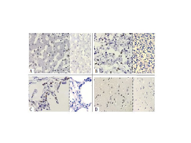
Validation of chromogenic in situ hybridization reactions for detection of DNA and RNA in formalin-fixed and paraffin-embedded tissues
Rachel L. Monteiro; Daniela S. Damaceno; Lidia M. Kimura; Cinthya S. Cirqueira; Juliana M. Guerra; Leonardo José T. AraújoJ. Bras. Pathol. Med. Lab. 2019;55(1):49-58doi.org/10.5935/1676-2444.20190008 ABSTRACT INTRODUCTION: Chromogenic in situ hybridization (CISH) is used alternatively to the traditional immunohistochemical methods for the diagnosis of infectious diseases in formalin-fixed paraffin-embedded samples, since it presents high sensitivity and specificity. This type […]